
For the 4th consecutive year, the European Dry Eye Society (EuDES) organised a photo contest open to all its EuDES members. The winner received a full invitation for the EuDEC 2026 in Milan on June 18-20, 2026 and the best pictures were displayed during the congress.
Discover the best pictures of the 2026 contest
Click to show the captions
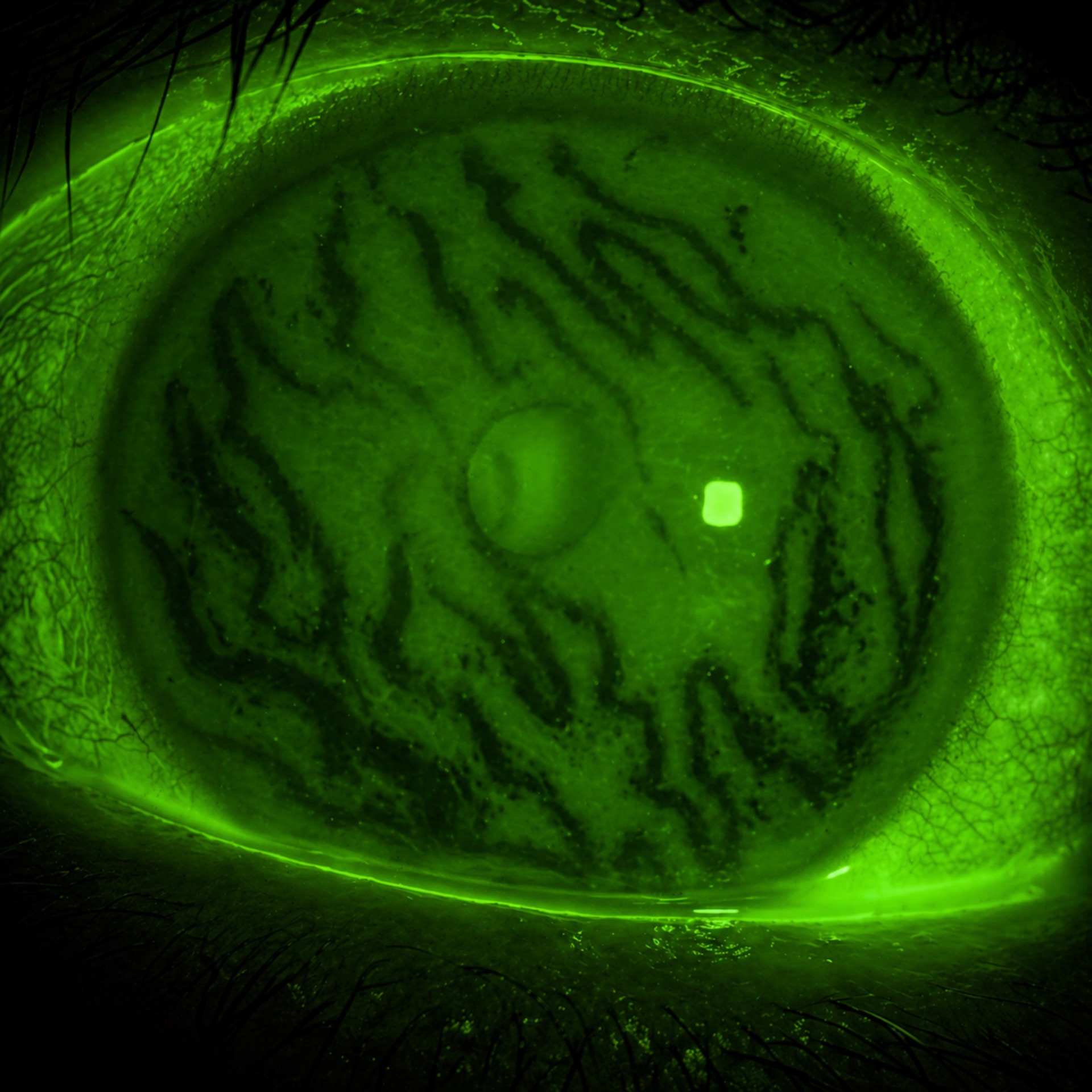
#1st price : Emerald Silence
University of Seville · Seville, Spain
In the green hush of light, the eye becomes a hidden world - fragile, luminous and full of untold tides.
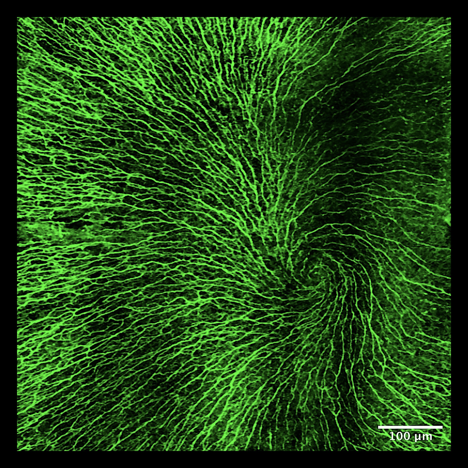
#2nd ex-aequo price : Spiral of Sensation
Instituto de Oftalmologia FAP Conde de Valenciana, IAP · Mexico, Mexico
This image reveals the vortex-like organization of the corneal subbasal nerve plexus, a dynamic network essential for sensation, tear regulation, and epithelial integrity. In dry eye disease, disruption of thisdelicate architecture reflects impaired sensory feedback, tear film instability, and ocular surface damage.
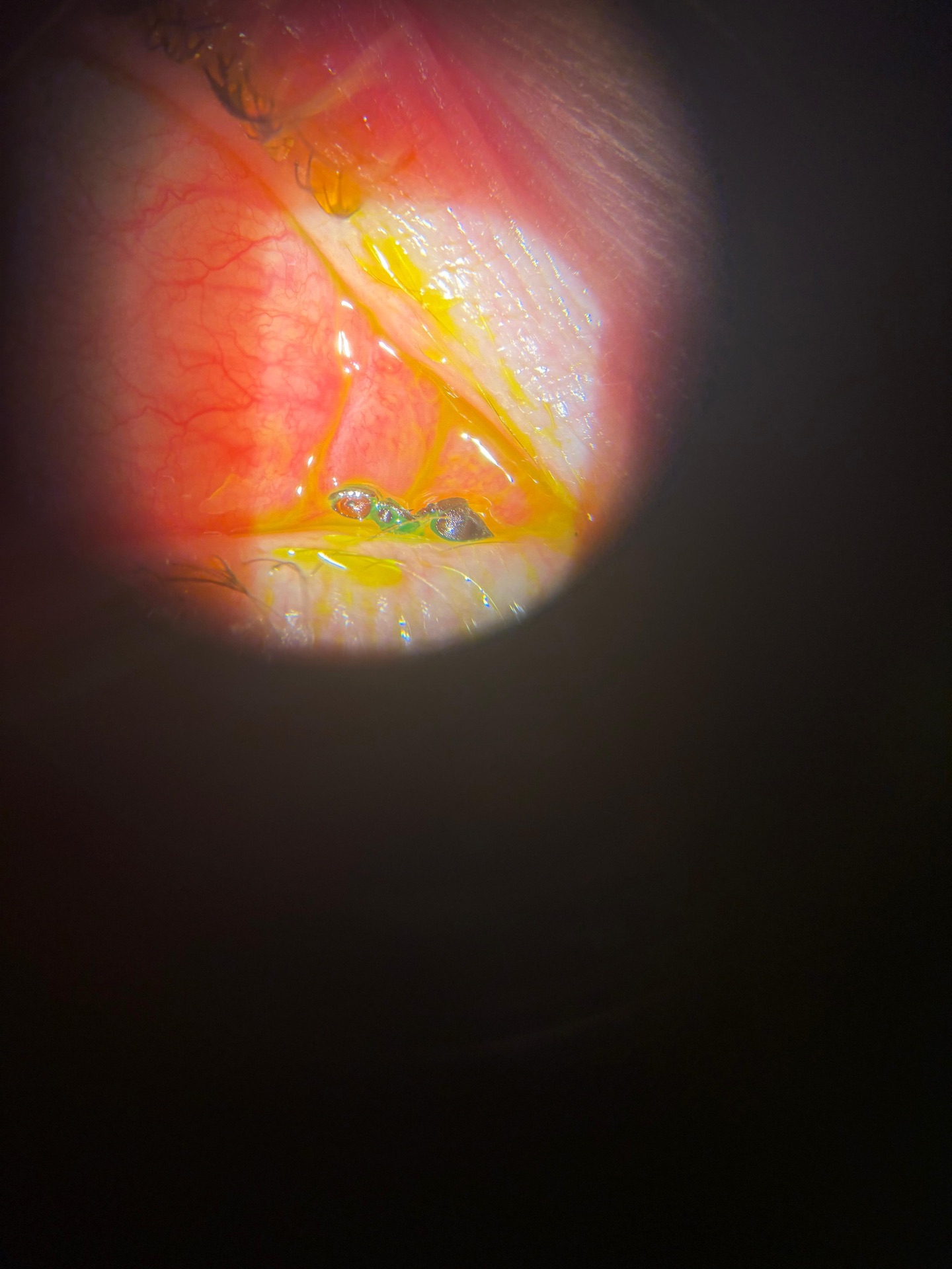
Unexpected Foreign Body : Ant at the Ocular Surface
Department of ophtalmology, arnas civico hospital · Palermo, Italy — Team: Gregorio Lo Giudice & Claudio Brancato
A 30 year old woman presented to the emergency eye department at night with acute burning and foreign bidy sensation. Slit-lamp examination revealed a dead red ant firmly attached to the medial canthus. While the body was removed with forceps, the head remained achored to the conjunctival tissue, requiring delicate dissection with a 15° blade. This unusual case highlights how even a tiny intruder can cause significant ocular discomfort and pose unexpected removal challenges.
BECOME A MEMBER FOR FREE
Contact us |