The European Dry Eye Society (EuDES), within the framework of the EuDEC 2024 in Madrid on June 20-22, 2024, organises a photo contest open to all EuDES members.
The best picture award will include a full invitation for the EuDEC 2024 in Madrid (travel, one night accommodation, and free registration).
So, make sure to send your best picture by May 15, 2024 along with the competition entry form to support-eudec@europa-organisation.com
Discover the best pictures of the 2023 contest:
Click to show the captions
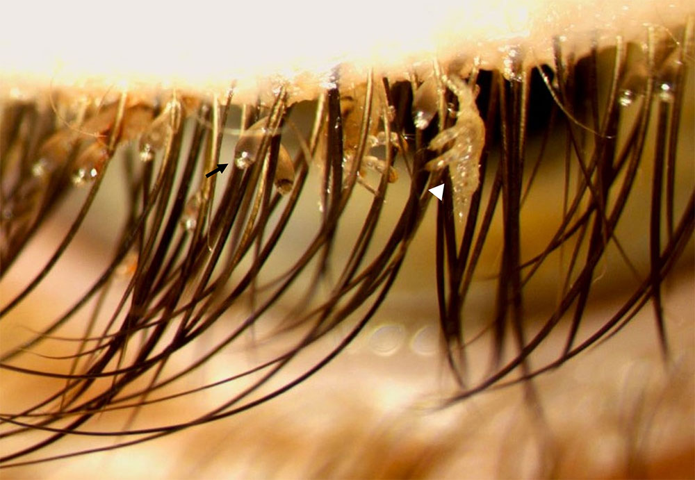
"Hang On"
A 5-year-old female presented to the ophthalmology department with complains of ocular pruritus for the past month. Slit-lamp biomicroscopy revealed translucent egg-shaped nits (black arrow) and adult lices (white arrowhead) attached to the eyelashes. Microbiological examination identified Pthirus pubis.
These infestations may spread through close contact with an infected individual and sexual abuse must be excluded as this can be a sexually transmitted disease. She was referred to the dermatology and paediatric departments to treat associated infestations and rule out other sexually transmitted diseases.
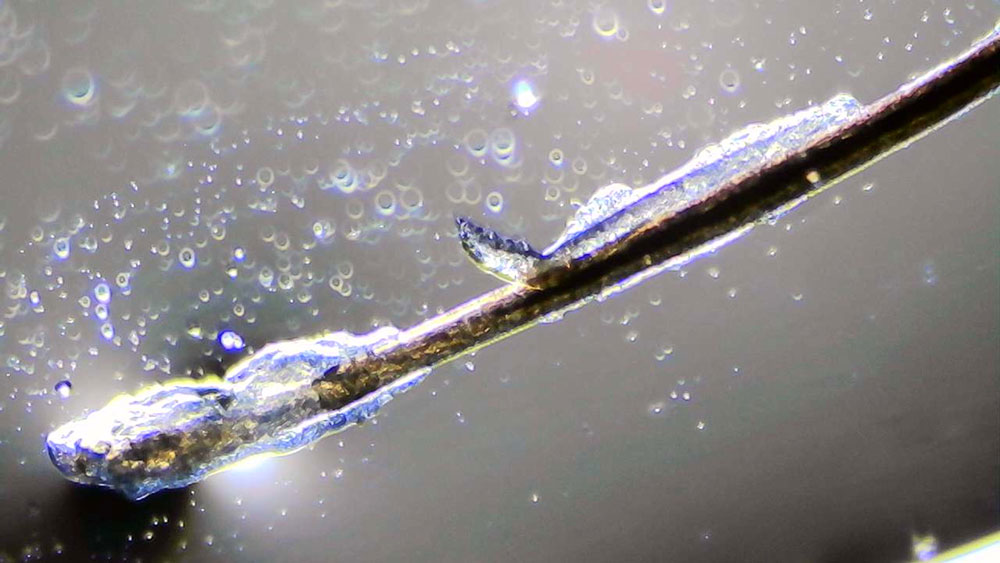
"A Resting Demodex"
After epilation of an eye lash, we put it on a microscope slide with immersion oil. Surprisingly oil and the backlight formed this magnificient view of a demodex.
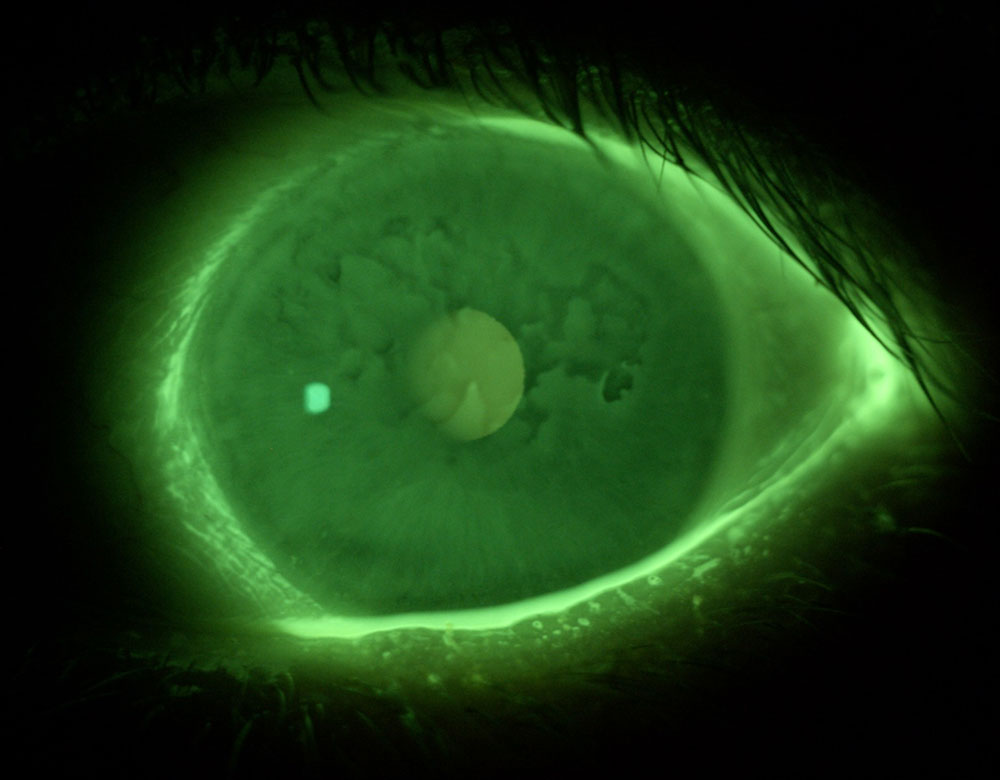
"Mixture of Corneal Dystrophies on Ocular Surface"
Coexisting Epithelial Membrane Corneal Dystrophy and Fuchs Endothelial Corneal Dystrophy. The photo was taken after fluorescein installation with blue-yellow filters.
BECOME A MEMBER FOR FREE
Contact us |